FIG 6.
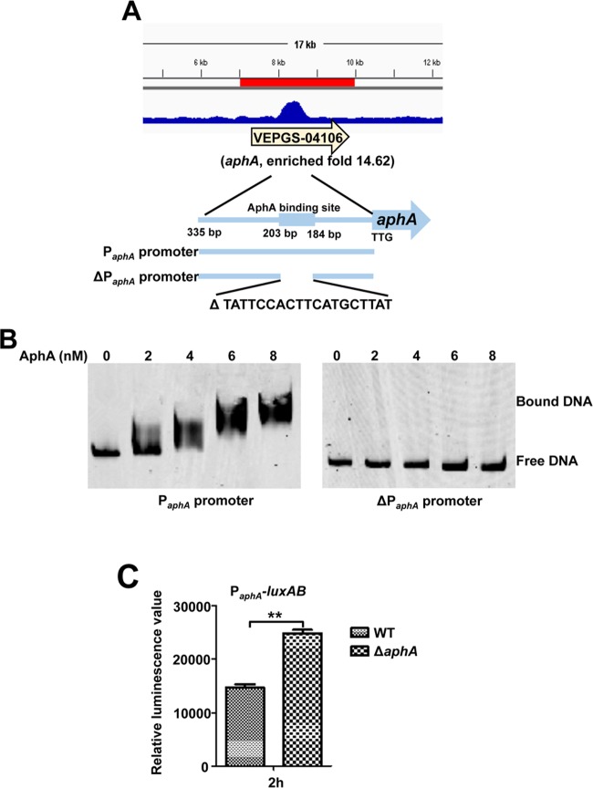
AphA binds directly to its own promoter for autorepression. (A) AphA binds to its own promoter region as illustrated with the peak (enriched 14.6-fold) identified by the ChIP-seq experiment. The AphA binding site and the probes (335-bp PaphA promoter and 316-bp ΔPaphA promoter with deletion of AphA binding site ranging from bp 184 to 203 relative to start site TTG) used for EMSAs in panel B are illustrated. (B) EMSA of the aphA promoter region (left) or the aphA promoter region with deletion of the specific AphA-binding box (ΔPaphA) (right) with purified AphA. The amounts of AphA protein used are indicated, and 20 ng of each Cy5-labeled probe was added to the EMSA mixtures. The specificity of the shifts was verified by adding a 10-fold excess of nonspecific competitor DNA [poly(dI·dC)] to the EMSA mixtures. (C) PaphA-luxAB transcriptional analysis in WT and ΔaphA strains. The WT and ΔaphA strain carrying the PaphA-luxAB reporter plasmid were cultured in LBS for 2 h and assayed for luminescence. The results are shown as means ± SD (n = 3). **, P < 0.01 (Student's t test) compared to the value for the WT strain harboring PaphA-luxAB.

