Fig. 2.
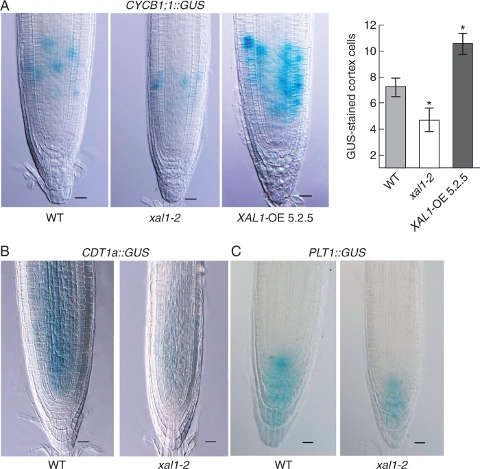
XAL1 positively regulates CYCB1, CDT1 and PLT1 in the RAM. (A) Lower and higher levels of pCYCB1;1::GUS expression in xal1-2 roots and XAL1-OE 5.2.5 respectively, compared with WT. The number of GUS-stained cortex cells is shown on the right. Data correspond to mean ± s.e. and statistically significant (*) differences from WT (P < 0·05) were determined with the Kruskal–Wallis test. (B, C) Expression of pCDT1a::GUS (B) and PLT1::GUS (C) is diminished in xal1-2 roots compared with WT roots. All plants are 5 dpg, n = 10 per line. Scale bars = 20 µm.

