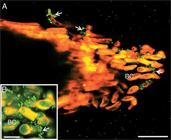
Fig. 6.

Laser confocal microscopy image showing binding of the bacterium P. atrosepticum to S. tuberosum root tip. Note the presence of bacteria over border cells (arrowheads and inset). Green spots correspond to bacteria. BC, border cells. Scale bars: (A) = 86 µm (A); (B) = 20 µm.
