
Figure 2.
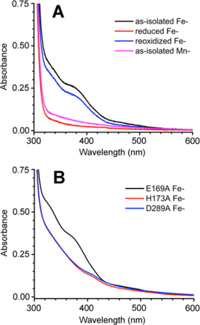
UV–vis absorption spectra of (A) Fe- and Mn-TM0186 and (B) E169A and H173A Fe-TM0186. Proteins were at a concentration of 50 μM in 50 mM Tris, 50 mM NaCl, and 10 mM MgCl2 (pH 7.6). The weak absorption feature near 410 nm in the H173A and D289A Fe-TM0186 spectra is due to a very small amount of contaminating heme.