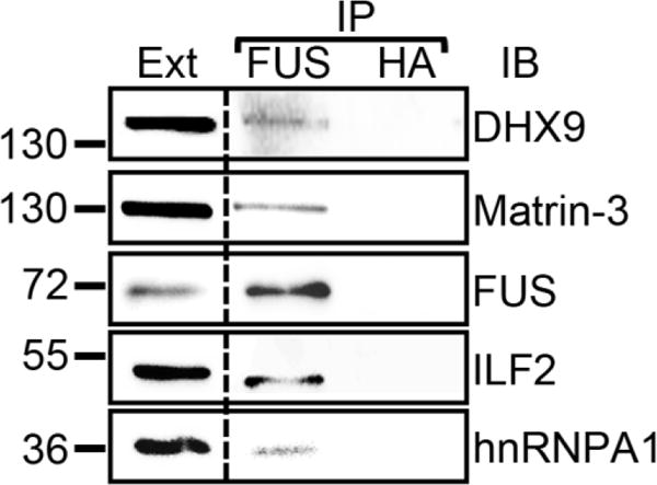
Figure 2. Verification of selected FUS interacting partners with endogenous FUS immunoprecipitation.

Immunoprecipitations were performed from cellular extracts of SH-SY5Y cells with a FUS-specific and a control (HA) antibody. The immunoprecipitates were subjected to SDS-PAGE followed by immunoblot with the indicated antibodies. IP, immunoprecipitation; Ext, extract; IB, immunoblot. The molecular weights of nearby marker bands are shown on the left (kDa).
