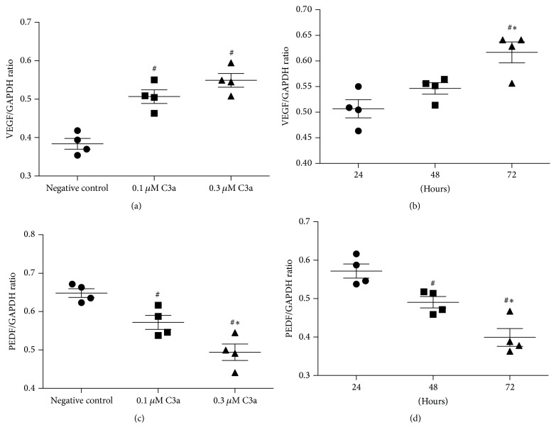
Figure 1.
Effect of exogenous C3a on the levels of VEGF and PEDF mRNA in cultured ARPE-19 cells. Total RNA was extracted and mRNA evaluated by RT-PCR analysis. The ratio of the abundance of each mRNA to that of GAPDH was evaluated by densitometric analysis. Data are mean ± SD (n = 4). (a) and (c) show the levels of VEGF and PEDF mRNA incubated with different doses of C3a for 24 hours; # P < 0.05 versus negative control cells; ∗ P < 0.05 versus 0.1 μM C3a incubation; (b) and (d) show the levels of VEGF and PEDF mRNA incubated with 0.1 μM C3a for 24, 48, and 72 hours. # P < 0.05 versus 24 hours of incubation; ∗ P < 0.05 versus 48 hours of incubation.

