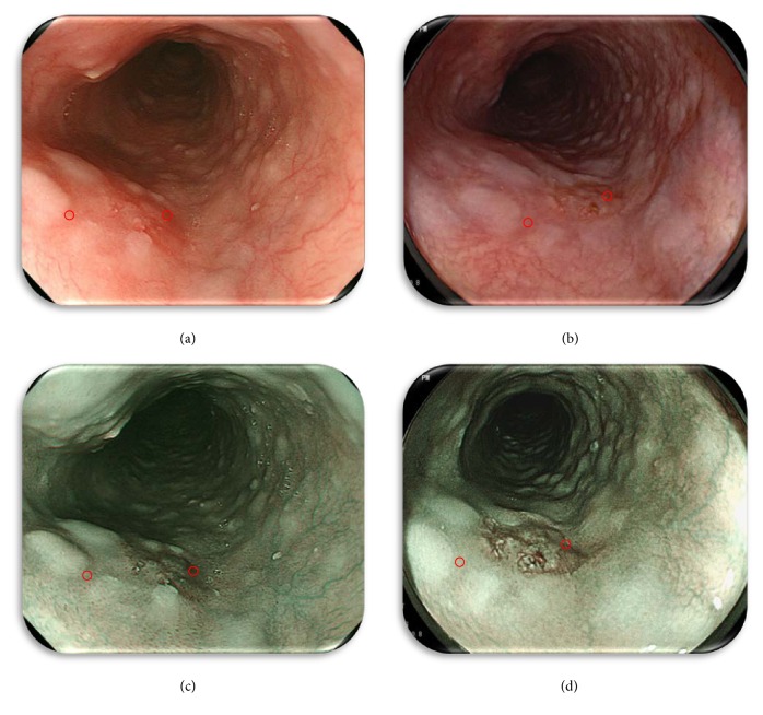
Figure 2.
Representative still images illustrating the spots captured for the color difference score (CDS) calculation of the lesion and background mucosa (Figure 1). (a) O-WLI image, (b) F-WLI image, (c) NBI image, and (d) BLI-bright image. The lesion was captured for image processing, and the region of interest (ROI) was highlighted to calculate the CDS using each of the four methods.

