Figure 3.
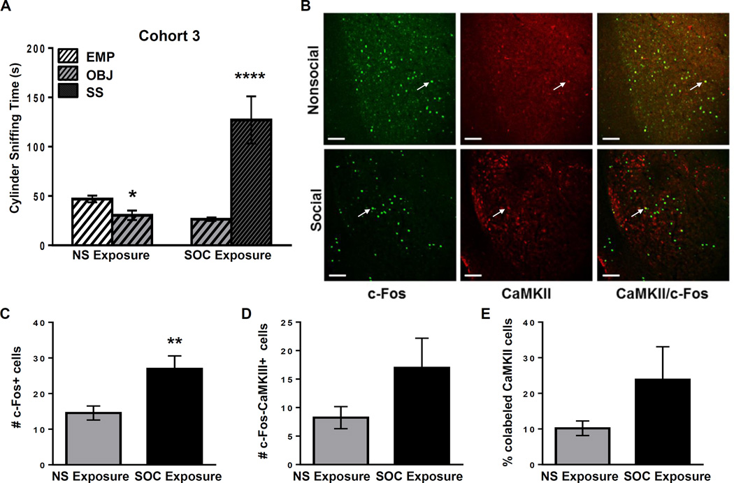
Cohort 3: B6 mice show high social approach and glutamatergic activation of the BLA. A: Cylinder sniffing times for the NS (n=6) and SOC (n=5) B6 mice in Cohort 3 in Phase 2 of the SAT. **** =p<0.0001 compared to OBJ cylinder. B: Representative double immunofluorescent staining in the BLA with c-Fos (green), CaMKIIα (red) and both (yellow) in Cohort 3 B6 juvenile males in nonsocial (NS) and social (SOC) exposure groups. White arrows indicate representative co-labeled cells. Scale bars = 100 µm. C: Number of c-Fos+ cells in the BLA of B6 NS or SOC mice. ** = p<0.010. D: Number of BLA cells colabeled with c-Fos and CaMKII. E: Percent of CaMKII+ BLA cells that are co-labeled with c-Fos in the NS or SOC exposure groups. Raw data is presented in the graphs, but a log transformation was required for normality to perform data analysis.

