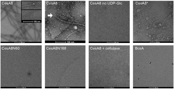
Fig. 7.
PttCesA8 produces cellulose microfibrils in vitro. Cellulose synthesized by reconstituted PttCesA8 was visualized by negative-stain EM. Shown are representative images of cellulosic material formed by the indicated PttCesA8 constructs. Cellulose microfibrils are seen and in some cases are associated with globular particles that may be cellulose synthase complexes. Cellulose microfibrils originating from a membrane sheet are indicated by a white arrow. Negative controls in the absence of UDP-Glc or with the inactive PttCesA8* mutant result in no fiber formation. Infrequent fiber formation is seen with the N-terminal truncation mutants. Treatment of the sample with cellulase for 10 min before grid preparation results in the loss of cellulose fibers. BcsA-produced cellulose was not detectable.

