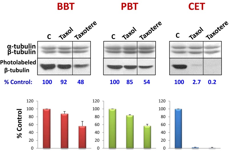
Fig. S2.
Inhibition of photolabeling by Taxotere. Four micromolar BBT, PBT, and CET were incubated with 5 µM [3H]2-m-AzTax in the absence or presence of fourfold molar excess (20 µM) Taxol or Taxotere. Samples were UV-irradiated and analyzed by SDS/PAGE as described in Materials and Methods. The bands in the upper panels represent α- and β-tubulin stained with Coomassie Blue. Photolabeling data were plotted as %Control ± SE, from 2 independent experiments.

