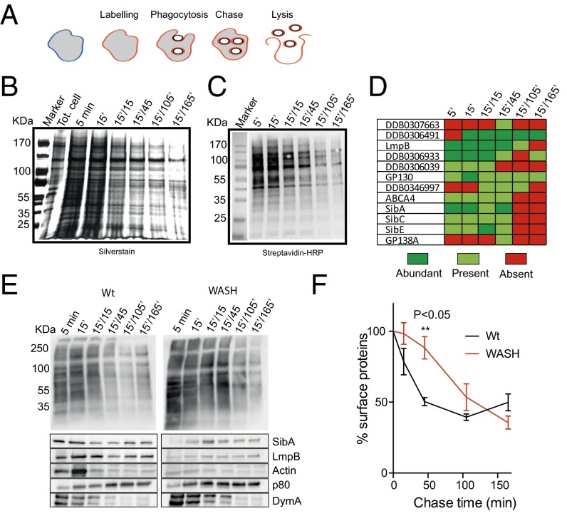
Fig. 7.
Internalization of surface proteins by phagocytosis. (A) Outline of experimental procedure. The cell surface was labeled by biotinylation, before cells were allowed to phagocytose latex beads for 5 or 15 min. Beads were then washed out and phagosomes isolated at different time points, and surface-originated biotinylated proteins purified. These were analyzed by both (B) silver staining and (C) streptavidin Western blot. (D) Transmembrane proteins identified from each sample by mass spectrometry. Relative abundances were semiquantitatively assessed by number of peptides identified (n = 2). (E) Comparison of phagosome maturation in wild-type and WASH-null cells. Surface labeling and phagosome purification was done in parallel and probed with either streptavidin (Top) or specific antibodies (Bottom). (F) Quantification of biotinylated proteins on purified phagosomes, by densitometry of the entire lanes in blots such as in E (n = 2). All times indicated are in the format:pulse time (min)/chase time (min).

