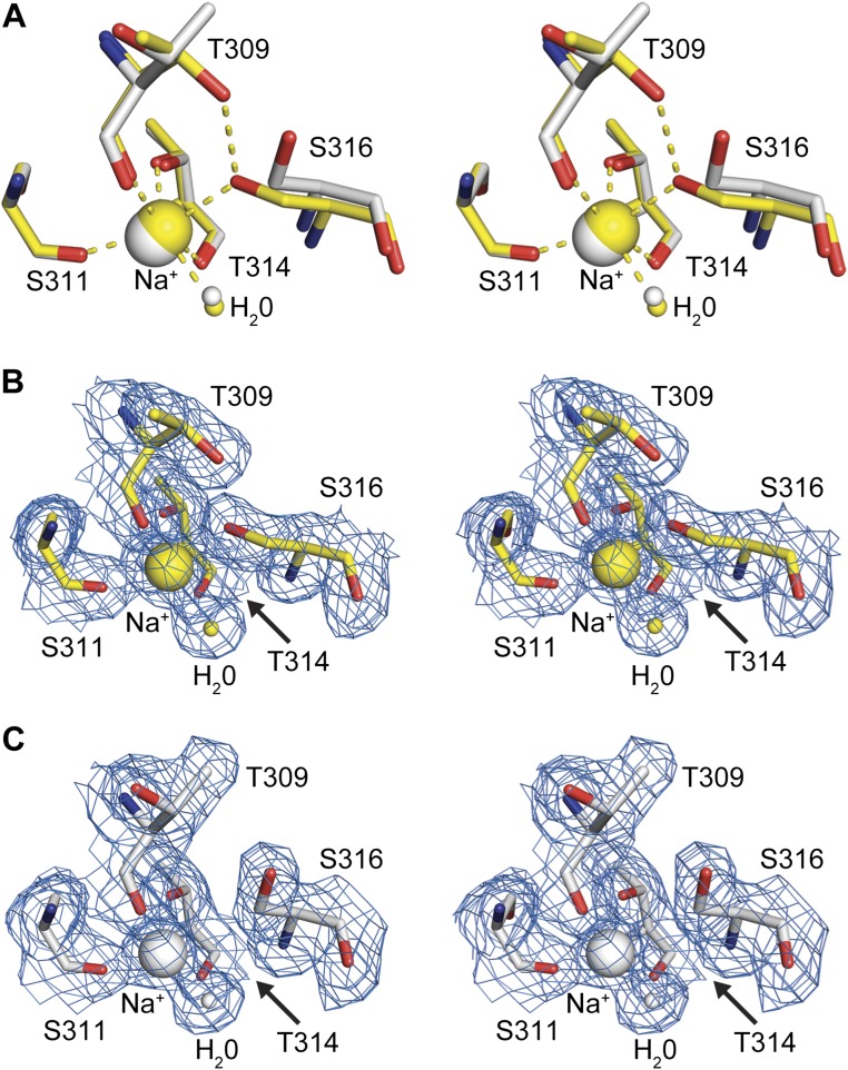
Fig. S5.
Substrate-induced conformational changes of the sodium site in furin. Stereo panels show a superposition of selected residues of unliganded furin (yellow, protein: stick model; nonbonded atoms: spheres) and inhibitor-bound furin (gray, protein: stick model; inhibitor: ball-and-stick; nonbonded atoms: spheres). (A) Superposition of the unliganded inhibitor-bound states. (B and C) Electron-density maps observed for the sodium binding site. Sodium ions and water molecules are given as big and small spheres, respectively. The 2Fo–Fc simulated annealing composite-omit electron-density maps are given as blue-colored mesh and are contoured at 1.0 σ. (B) Unliganded furin. (C) Furin complexed with MI-52.

