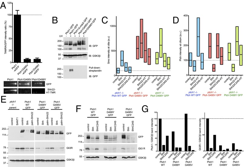
Fig. 6.
Defective palmitate-dependent interaction with Shh in Ptch1 mutants causing Gorlin Syndrome. (A) Binding of Shh22-HT-TMR to cilia in Ptch1-null cells rescued with eGFP-tagged Ptch1, Ptch1G495V, or Ptch1D499Y was measured by live imaging. Graph shows ratio of TMR and GFP fluorescence at cilia. Error bars represent SE (n > 14 cilia). Representative images of cilia are shown below the graph. Ptch1G495V and Ptch1D499Y do not bind Shh22-HT-TMR. (B) Ptch1-null cells, stably expressing the indicated Ptch1 constructs, were incubated with 15-azi-palm-Shh22-biotin (3.5 μM). The cells were UV-irradiated, and photocrosslinking was analyzed by denaturing affinity precipitation with streptavidin, followed by SDS/PAGE and immunoblotting. GSK3β was used as loading control. Ptch1G495V and Ptch1D499Y are not photocrosslinked to 15-azi-palm-Shh22-biotin, in contrast to Ptch1. Ptch1Δloop2, which does not bind palm-Shh22, serves as negative control. (C) Ptch1-null cells rescued with eGFP-tagged Ptch1, Ptch1G495V, or Ptch1D499Y were incubated with control media, Shh, Shh22-HT, or Shh22C24S-HT, and endogenous Smo recruitment to cilia was measured by immunofluorescence and automated image analysis (n > 300 cilia). Ptch1G495V and Ptch1D499Y are impaired in their response to Shh22-HT but respond normally to Shh. (D) As in C, but showing Ptch1 localization at cilia. (E) As in C, but cells were treated with control media, Shh, or palm-Shh22 (5 μM), and Gli3R, Ptch1, and GSK3β were analyzed by immunoblotting. In cells expressing Ptch1G495V and Ptch1D499Y, Gli3R levels decrease in response to Shh but not to palm-Shh22. (F) As in E, but cells were treated with control media, Shh, or ShhC24S. Cells expressing Ptch1G495V and Ptch1D499Y respond to both Shh and ShhC24S, whereas Ptch1 responds preferentially to Shh. (G) Quantification of the experiment in F.

