Fig. 3.
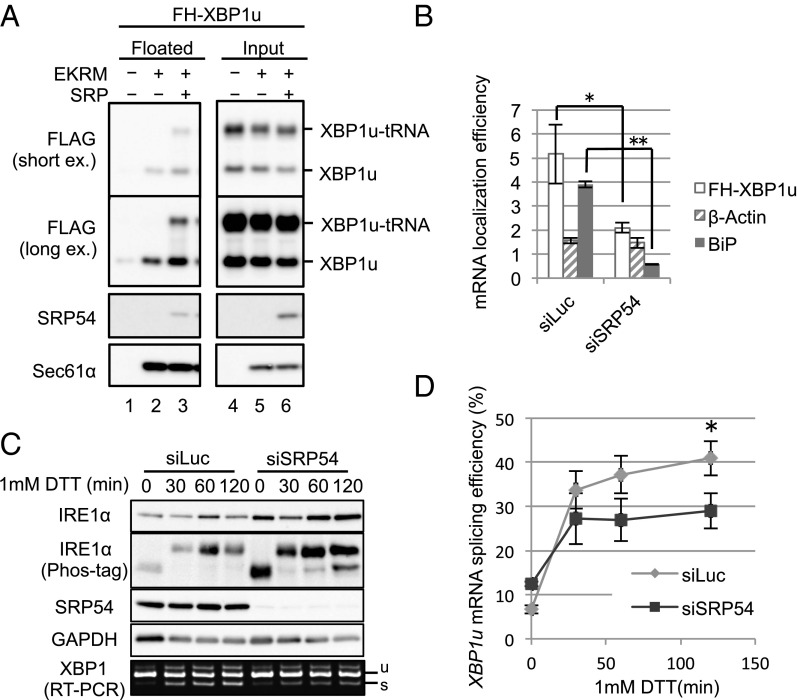
SRP recruits XBP1u-RNC to the ER. (A) FH-XBP1u was translated in WGE in the presence or absence of 5 nM purified SRP and CMMs treated with EDTA and high-salt medium (EKRM) to remove preexisting SRP on CMMs. The membrane-bound proteins were separated by a membrane-flotation assay. Proteins in those fractions were detected by immunoblotting. (Top and Middle Top) Same result of immunoblotting exposed for a short time and a long time, respectively, is shown. (B) Membrane localization efficiency (membrane-bound/cytosol) of XBP1u, β-actin, and BiP mRNA in HeLa cells stably expressing FH-XBP1u with SRP54 knockdown for 96 h was quantified as described in Fig. 2C. Bars indicate SD. *P < 0.05, **P < 0.01 (n = 3) in siLuc (Control) vs. siSRP54 using Student’s t test. (C) HeLa cells stably expressing XBP1u-ps with SRP54 knockdown were treated with 1 mM DTT for the indicated times. Western blot analyses of the phosphorylated state of IRE1α and the abundance of indicated proteins are shown. The splicing of XBP1u-ps mRNA was analyzed by RT-PCR. (D) Proportion of the spliced form with respect to the total XBP1u-ps mRNA was calculated from C. Bars indicate SD. *P < 0.05 (n = 3) in siLuc (Control) vs. siSRP54 at 120 min using Student’s t test.

