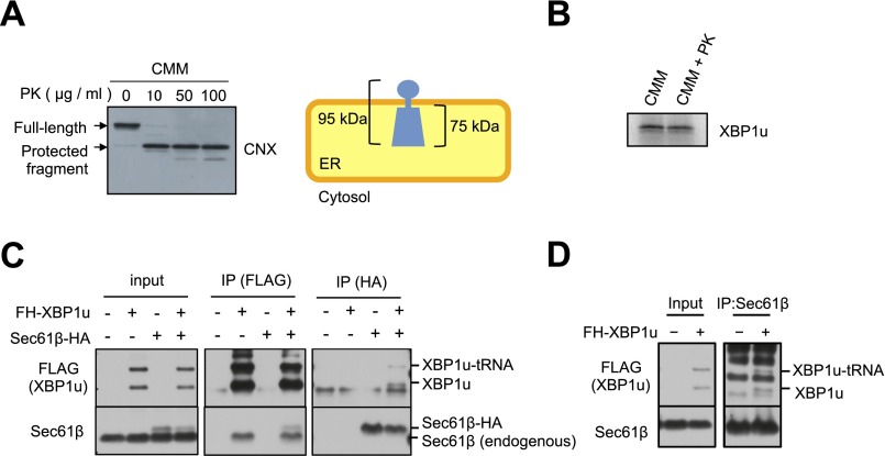
Fig. S2.
Supporting information for Fig. 2. (A) Preparation of proteinase K (PK)-treated microsomes. CMMs were treated with the indicated concentration of PK. The efficiency of surface protein digestion was evaluated by the removal of a short cytosolic tail of calnexin. (Right) Membrane topology of calnexin is shown. The full-length calnexin and protected fragment of calnexin were detected using the antibody against the ER-luminal portion of calnexin. (B) XBP1u translation was not affected by PK treatment in the presence of a PK inhibitor. XBP1u was synthesized via the in vitro translation system with mock-treated CMMs or PK-treated CMMs with PMSF (2 mM) and benzamidine (2 mM). (C) Endogenous Sec61β and/or HA-tagged Sec61β was coimmunoprecipitated with FH-XBP1u and FH-XBP1u-tRNA using anti-FLAG antibodies. In contrast, FH-XBP1u and/or FH-XBP1u-tRNA was coimmunoprecipitated with Sec61β and/or Sec61β-HA from the cell lysate derived from HEK293T cells transiently expressing FH-XBP1u and/or Sec61β-HA. Anti-FLAG or anti-HA antibody was used for immunoprecipitation (IP). (D) Coimmunoprecipitation of endogenous Sec61β with FH-XBP1u from the cell lysate derived from HEK293T cells transiently expressing FH-XBP1u. Anti-Sec61β antibody was used for immunoprecipitation.

