Fig. S3.
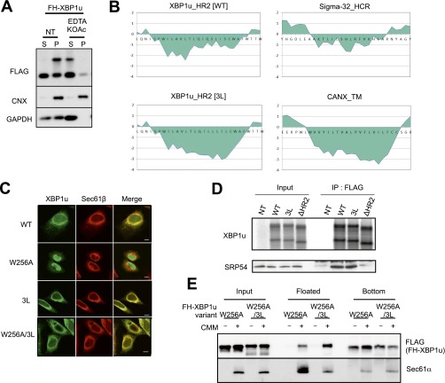
Supporting information for Fig. 5. (A) EDTA and high-salt treatment of microsomes in HEK293T cells transiently expressing wild-type or 3L mutant (3L) FH-XBP1u were analyzed by Western blotting. Calnexin (CNX) and GAPDH were used as the control membrane and cytosolic protein, respectively. (B) Kyte and Doolittle hydrophobicity plots for HR2 of WT and 3L mutant XBP1u (window size: 9) are visualized using a ProtScale analysis implemented in ExPASy (web.expasy.org/protscale/). (C) Variants of FH-XBP1u transiently expressed in HeLa cells were costained with Sec61β. (Scale bars, 10 μm.) (D) FH-XBP1u variants translated with RRL were coimmunoprecipitated with SRP54. XBP1u was detected by autoradiography, whereas SRP54 was detected by immunoblotting. (E) Posttranslational ER-targeting assay of FH-XBP1u[W256A] and FH-XBP1u[W256A/3L]. Total protein before fractionation is shown as the input. Floated and bottom fractions are shown. The presence and absence of the ER membrane (EKRM) are indicated by “−” and “+”, respectively. Additional information is provided in Materials and Methods.

