Fig. 3.
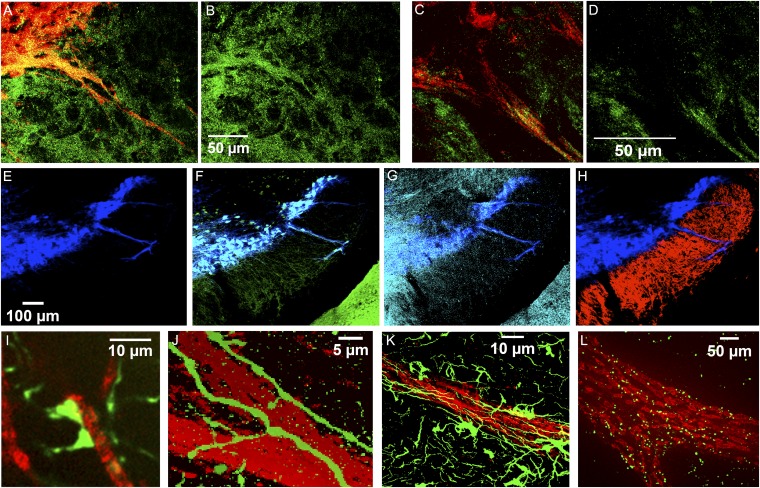
Immunomarkers identify multiple afferents within striosome–dendron bouquets. (A–D) Merged images of DAT-immunolabeled dendrons (red) coimmunolabeled for GAD65/67 (A and B; n = 10 sections from 5 animals) or VGluT2 (C and D; n = 10 sections from 2 animals) (green). (E–H) DAT-immunolabeled dendron (blue, E) coimmunolabeled for D2-GFP (green, F) and μ-opioid receptor (cyan, G) and weakly labeled with D1-tdTomato (red, H) (n = 3 sections from 1 animal). Similar results were obtained with a related line and by ProExM (Fig. S3). The edge of the cerebral peduncle is visible at the lower right corners in F and G and is immunolabeled for D2-GFP and μ-opioid receptor. (I) With ProExM, two mCitrine-positive (green) presumed contacts onto a DAT-positive (red) fiber are visible in the P172 striosome line (n = 2 sections from 1 animal). (J–L) Single-plane confocal images at the centers of dendrons (immunolabeled for DAT in red) showing cholinergic fibers (green) aligned along and within the dendron in the ChAT-Cre knock-in line with the Ai32-YFP reporter (J; n = 4 sections from 1 animal; see also Figs. S4 and S5), astrocytes (green, immunolabeled for GFAP) associated with dendron (K; n = 6 sections from 2 animals; see also Fig. S6 A–C), and ProExM of connexin-43 (green) junctions along the dendron (L; n = one section from 1 animal; see also Fig. S6 D–F, n = 4 sections from 1 animal). (Scale bars in B, D, E, J, and K indicate the dimensions of unexpanded tissue; scale bars in I and L indicate the dimensions of the expanded tissue.)

