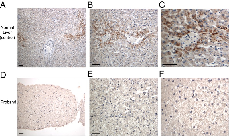
Fig. 2.
Absence of ACOX2 in the proband’s liver. (A–C) Immunohistochemistry for ACOX2 in normal liver shows intense granular staining in the pericentral (zone 3) hepatocytes, but faint staining in remaining hepatocytes, including periportal hepatocytes (zone 1). (D–F) Immunohistochemistry for ACOX2 in the proband’s liver biopsy, showing complete absence of staining. (Scale bars: 50 μm.)

