Table 1.
Microphysiological Technologies Used to Model the Hepatic Microenvironment
| Representative Image | Platform | Scaffold/Format | Co-culture or integrated with other organ tissues |
Validated functions of phase I/II and transporters |
Other liver-specific functions |
Methods used to assess hepatotoxicity |
|---|---|---|---|---|---|---|

|
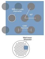
HepatoPac Hepregen (32-33) |
Micropatterned 24- and 96-well plates |
Primary hepatocytes islands co-cultured with 3T3 fibroblasts |
Metabolite measurements:
CYP1A: Ethoxy-resorufin; CYP2A6: Coumarin; CYP2B6: Bupropion; CYP3A4: testosterone UGTs and SULTS: 7- hydroxycoumarin mRNA expression : phase I CYPs, FMO, MAO, OX, EH; phase II- UGTs, SULTs, NATs, GSTs, methyltransferases Transporters-MDR1, MRP3, OCT1, NTCP, BSEP canalicular flow by CMFDA fluorescent probe |
Albumin (ELISA) and urea (colorimetric endpoint assay) secretion |
MTT assay, ATP, and glutathione levels using Cell Titer-Glo and GSH-Glo luminescent kits (Promega) |

|
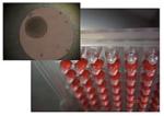
RegeneMed Liver Transwell RegenMed (35) |
24-well transwell plate | Primary human or rat hepatocytes with hepatic NPCs,including vascular and bile duct endothelial cells, Kupffer cells and hepatic stellate cells |
Phase I activity: CYP1A1, 2C9, and 3A4 by P450-Glo assays (Promega) Basolateral transporter uptake measured by isotope labeled tracer - 3H-labeled estrone-3-sulphate |
Albumin, urea, fibrinogen, transferrin production (ELISA); glycogen synthesis (isotope labeling) LPS induced IL-1β, IL- 6, IL-8, TNF-α, GM- CSF: multiplex-based ELISA |
Levels of ATP and GSH, LDH releasing |

|
3D InSight™ Microtissues InSphero (36) |
Spheroids in 96-well plate |
Primary human hepatocytes with NPCs (Kupffer and endothelial cells) |
CYPs activity/induction: CYP1A1, 2B6, 2C8,2C9,2C19,2D6 CYP3A4 and 2E1 (IHC staining) MDR1,BSEP (IHC staining) |
Albumin secretion (ELISA) IL-6 and TNF-α releasing (ELISA) |
ATP, and glutathione (GSH) levels using CellTiter-Glo and GSH-Glo luminescent kits (Promega) LDH releasing, mitochondrial activity |

|
3D print Liver Organovo |
3D bioprinted liver tissues in a transwell plate |
Primary human hepatocytes with NPCs |
CYP450 enzymes 3A4 (mRNA expression levels and probe metabolite formation) |
Albumin and transferrin production Immunologic: IL-6, GM-CSF,MCP-1 LPS stimulated TNF-α, IL-1β, IL-12p70, IL-10, IL-2, IL-13, and IL-4 |
LDH releasing, GSH and ATP level Gene/protein expression profiling and histological tissue assessment |

|
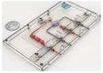
HμREL hepatic cultures Hurel (38) |
mCCA biochip connected with pump mCCA biochip can integrate multiple organ tissues |
Primary human hepatocytes with NPCs |
mRNA expression and
metabolite formation: CYP450 enzymes 1A2, 3A4, 2C19, SULT, UGT Metabolic clearance: Biliary efflux assay (LC-MS/MS) BSEP canaliculi (CMFDA) |
MTT assay, ATP, and glutathione (GSH) levels using CellTiter- Glo and GSH-Glo luminescent kits (Promega), LIVE/DEAD stain (Thermo Fisher) |
|

|
LiverChip™ System CN Bio Innovations (39-41) |
Multiwell plate platform with pneumatic pump |
Primary hepatocytes co- cultures with NPCs and Kupffer cells |
Metabolite formation:
CYP450 enzymes 1A2, 2B6, 2C9, 2D6, 3A4 |
LIVE/DEAD stain (Thermo Fisher) |
|

|
CellAsic (42) | Primary human hepatocytes |
Metabolite formation: CYP450 enzymes 1A1/2, 2C9, 3A4, UGT BSEP canaliculi (CMFDA) |
LIVE/DEAD stain( fluorescent probe; Thermo Fisher) |
||

|
SQL-SAL University of Pittsburgh (43) |
Nortis chip (Nortis,
Inc.) with microfluidic flow |
Primary human hepatocytes
with cell lines derived from NPCs |
Metabolite formation: CYP450
enzymes 1A, 2C9, 3A4 IHC staining: MDR1,MRP2, BSEP, BRCP |
TNF-α releasing |
LDH leakage,
fluorescent protein ROS biosensors Stellate cell activation and migration |

|
IdMOC™ In Vitro ADMET Lab (72) |
24 or 96-well plate format integrated with multiple organ |
Primary human hepatocytes with 3T3 cells |
MTT assay, ATP, and glutathione (GSH) levels Using CellTiter-Glo and GSH-Glo luminescent kits (Promega) |
||

|
Microcompartment hollow fiber BAL (37) |
Hollow fiber | Primary human liver cells ( not pure hepatocytes) |
Urea production , albumin
synthesis , glucose secretion , and lactate production IHC: expressed cell marker of Kupffer cells) |
LDH and AST releasing |
