
Figure 4. Widespread decreases in neocortical functional connectivity in focal epilepsy patients.
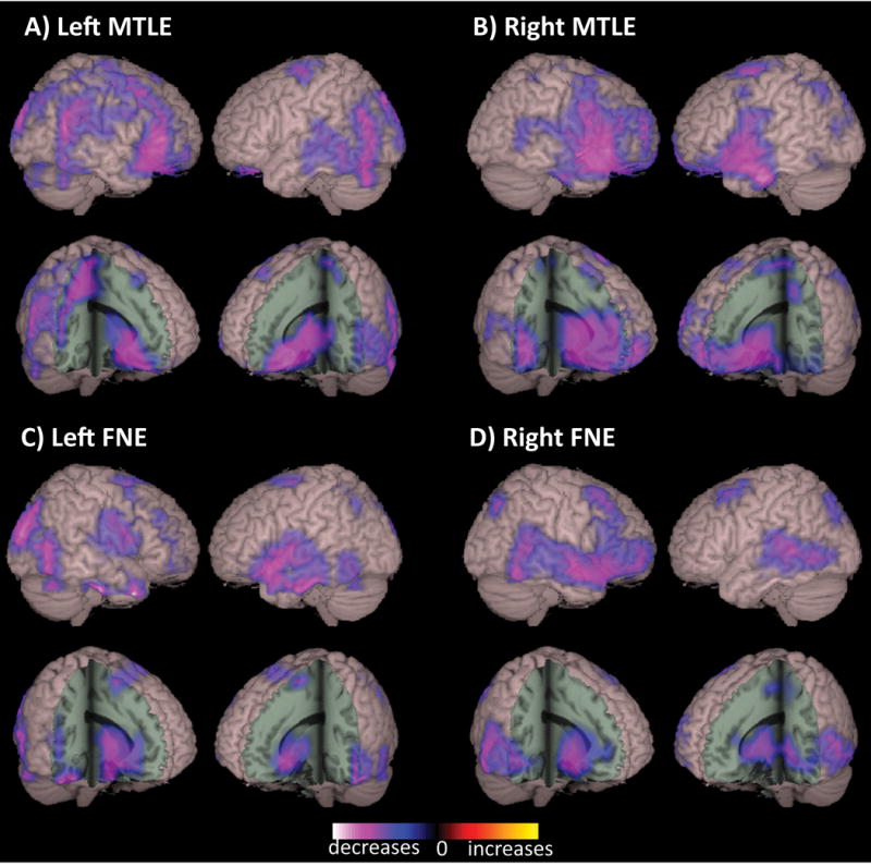
Compared to control subjects, patients with left MTLE (A), right MTLE (B), left FNE (C), and right FNE (D) demonstrated decreased functional connectivity in widespread neocortical regions, including fronto-parietal and posterior temporal association cortex, peri-sylvian neocortex, orbital frontal cortex, as well as decreased subcortical connectivity in basal forebrain and anterior thalamus. Connectivity maps represent t-tests (threshold p < 0.01, FDR-corrected) of alpha-band imaginary coherence in patients with left MTLE (N = 18), right MTLE (N = 12), left FNE (N = 17) or right FNE (N = 14) compared to controls, overlaid on a 3D-rendered template brain. FDR: false discovery rate; FNE: focal neocortical epilepsy; MTLE: mesial temporal lobe epilepsy; RSFC: resting-state functional connectivity. Modified with permission from Englot et al, 2015.43