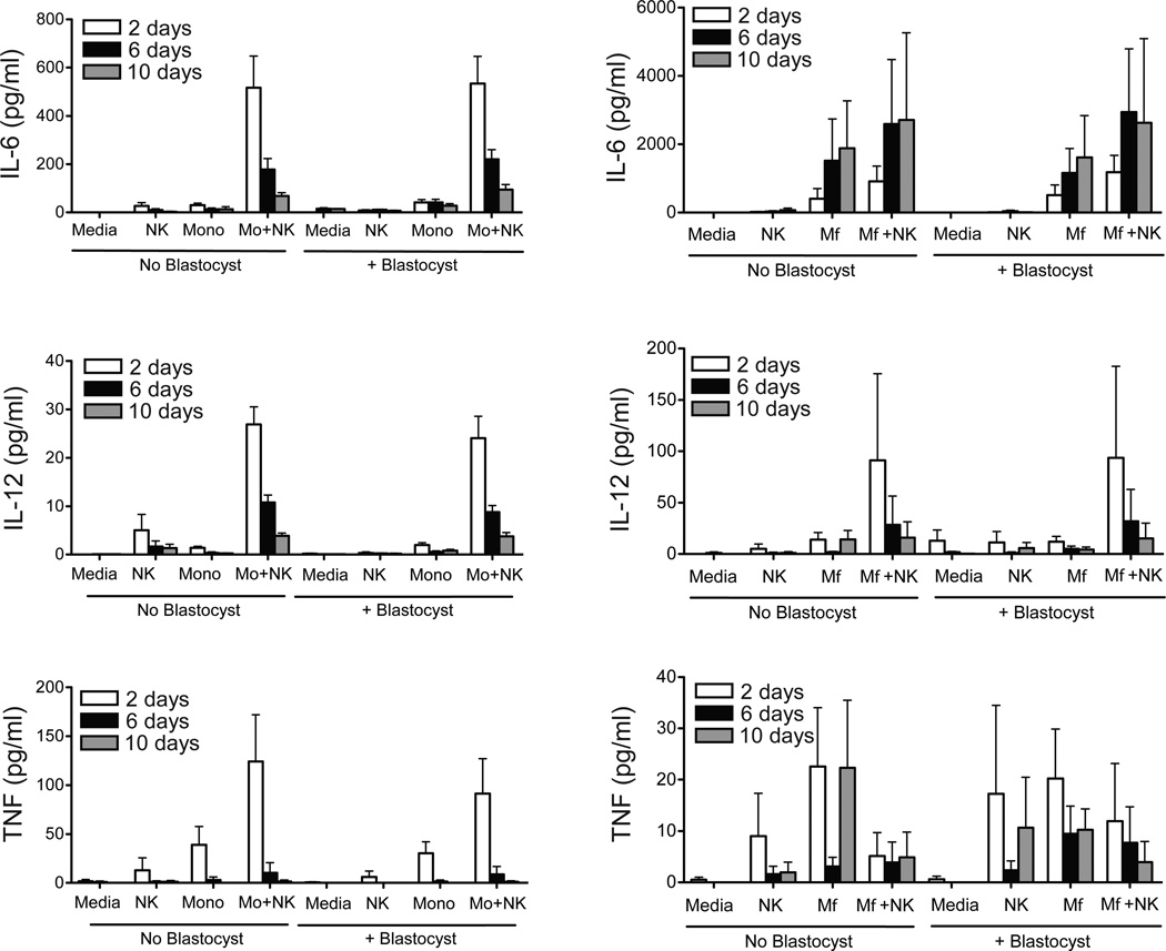
Figure 3.
IL-6, IL-12 (p40), and TNF concentrations in the media from leukocyte-blastocyst co-cultures on days 2, 6, and 10. The left panel represents levels within media for control wells with medium only (n=7), NK cells (n=8), monocytes (Mo, n=8), NK cells + monocytes (n=7) and in the right panels represent the levels within media for control wells with medium only (n=7), NK cells (n=8), macrophages (Mφ, n=8), or NK cells + Mφ (n=7). Data is represented as the mean ± SE. There were essentially no detectable levels of cytokines in media cultured in the absence of PBMCs.

