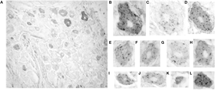
Figure 4.
Photomicrograph of 5-HT1F receptor immunoreactive neurons in the macaque trigeminal ganglia. (A) Notice the presence of considerably fewer immunopositive cells in general and fewer immunopositive large size cells in particular, compared to the vestibular ganglia. The calibration bar represents 40 microns; (B–L) Staining patterns and associated cell sizes in representative trigeminal ganglion cells. Most 5-HT1F receptor immunopositive trigeminal ganglia have a similar punctate staining pattern. A few large (B,D) and small (L) cells showed more intensely stained background cytoplasm.

