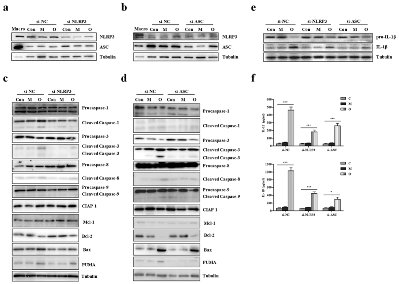
Figure 2. si-NLRP3 or si-ASC effect on the expressions of apoptosis associated proteins and levels of IL-1β and IL-18 in HK-2 cells responded to different kinds of contrast media.
(a) Western blot of NLRP3 and ASC in si-NC (non-specific control) and si-NLRP3. Macrophages are shown as positive controls (Macro). (b) Western blot of NLRP3 and ASC in si-NC (non-specific control) and si-ASC. Macrophages are shown as positive controls (Macro). (c) Western blot of procaspase-1,−3,−8,−9, cleaved caspase-1,−3,−8,−9, other apoptotic proteins (CIAP 1, Mcl-1, Bcl-2, Bax and PUMA) in si-NC (non-specific control) and si-NLRP3. (d) Western blot of procaspase-1,−3,−8,−9, cleaved caspase-1,−3,−8,−9, other apoptotic proteins (CIAP 1, Mcl-1, Bcl-2, Bax and PUMA) in si-NC and si-ASC. (e) Western blot of pro-IL-1β and IL-1β in si-NC, si-NLRP3 and si-ASC. (f) ELISA assay of the levels of IL-1β and IL-18. HK-2 cells transfected with si-NC or si-NLRP3 or si-ASC were treated with mannitol and omnipaque, respectively. Data are shown as mean ± SD. *P < 0.05, ***P < 0.001.

