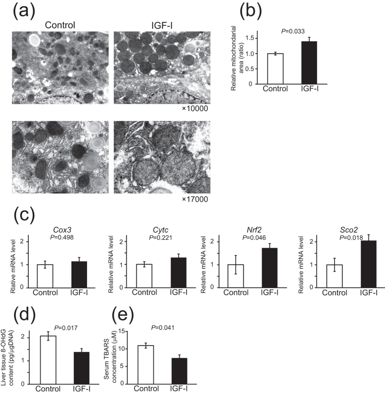
Figure 2. IGF-I improved the abnormalities in mitochondrial ultrastructure, mitochondrial gene expression, and oxidative stress markers in MCD-db/db NASH model mice.
(a) Ultrastructure assessed by electron microscopy (original magnification 10,000×and 17,000×) of the liver tissue. In the control liver, the size of the mitochondria was smaller and heterogenous, shape was irregular, and the mitochondria showed profound cristae disorganization and irregular shapes. In contrast, IGF-I-treated mice showed restored mitochondrial structure. (b) Quantitative analysis of mitochondrial area showed that the area was significantly increased in the mice treated with IGF-I than in controls (n = 3 for each groups). (c) Quantitative realtime PCR analysis of mitochondrial functional genes (n = 5 for each groups). (d,e) Quantitative analysis of oxidative stress markers (n = 5 for each groups). (d) 8-OHdG levels in the liver tissue. (e) Serum TBARs levels. Data were compared by Student’s t test.

