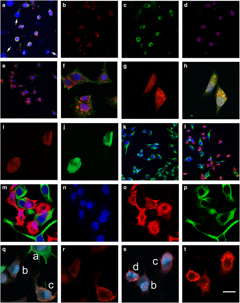
Figure 2.
MG63 cells were treated with cytotoxins for 19 h to induce PCD. Live staining with mAb2C4:AF568 and annexin V:AF488, was followed by fixation and staining with additional primary antibodies (caspase 8, LC3B or α-tubulin) followed by specific species or isotype secondary antibody:AlexaFluors. (a–d) MG63 cells were treated with 1 μM staurosporine: (a), a merged image, with arrows indicating examples of nuclei from unaffected cells; (b) mAb2C4:AF568; (c) annexin V:AF488; and (d) cleaved caspase 8. (e and f) merged images of MG63 cells were treated with 50 μM etoposide+30 μM necrostatin-1 (DAPI (4,6-diamidino-2-phenylindole), blue; mAb2C4:AF568, red; annexin V:AF488, green). (g and h) mAb2C4:AF568 (red channel) and merged image of MG63 cells treated with 50 μM chloroquine to induce autophagy (DAPI, blue; mAb2C4:AF568, red; LC3B, green). (i and j) MG63 cells treated with tumor necrosis factor α and zVAD-fms underwent necroptosis (mAb2C4:AF568, red; annexin V, green). (k) MG63 cells were untreated as controls (DAPI, blue; α-tubulin, green). (l–p) MG63 cells were treated with 1% DMSO+1 μM paclitaxel: (l, DAPI, blue; α-tubulin, green; mAb2C4:AF568, red) induced into the PACSR. (q–t) separate experiment with cells treated with 1% DMSO+1 μM paclitaxel: (DAPI, blue; mAb2C4:AF568, red; α-tubulin, pseudo-green) The calibration bar shown in (t) represents (a–e, k and l) 25 μm and (f–j, m-t) 10 μm.

