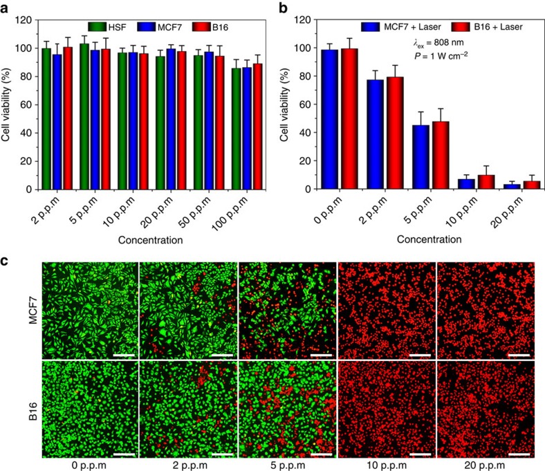
Figure 4. Cell experiments.
(a) Relative viability of the human skin fibroblast normal cells, MCF7 cancer cells and B16 melanoma cells after incubation with BPQDs/PLGA NSs (internal BPQDs concentrations of 0, 2, 5, 10, 20, 50 and 100 p.p.m.) for 48 h. (b) Relative viability of the MCF7 and B16 cells after incubation with BPQDs/PLGA NSs (internal BPQDs concentrations of 0, 2, 5, 10 and 20 p.p.m.) for 4 h after irradiation with the 808 nm laser (1 W cm−2) for 10 min. (c) Corresponding fluorescence images (scale bars, 100 μm for all panels) of the cells stained with calcein AM (live cells, green fluorescence) and PI (dead cells, red fluorescence).

