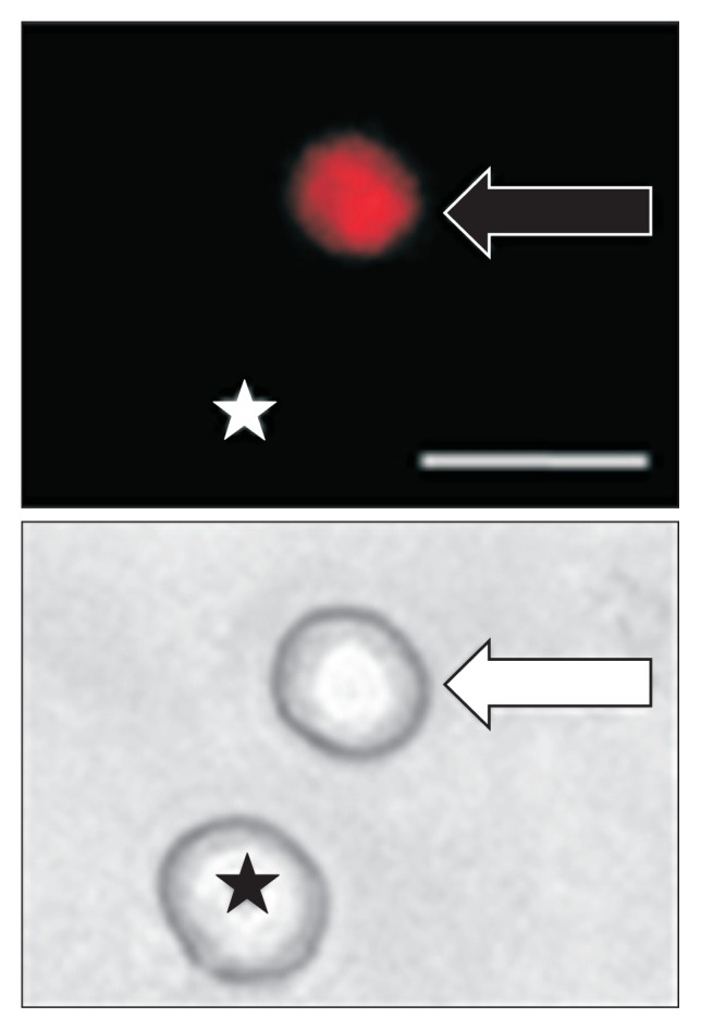
Figure 2.

Top: An example of a Dil-labeled dorsal root ganglion (DRG) neuron (arrow). Asterisk indicates the place where a neuron is not labeled by DiI. Bottom: Phase image of the same DRG neuron labeled by DiI is shown on the right (arrow) and the neuron not labeled by DiI is shown on the left (★). Scale bar = 50 μm. Patchclamp recordings were performed on DiI-labeled colon neurons. A total of 25 DiI-labeled neurons from control rats and 25 DiI-labeled neurons from streptozotocin-induced diabetic rats were recorded under current-clamp conditions.
