
Figure 2.
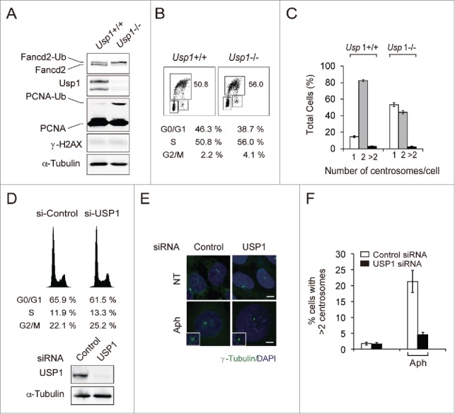
Usp1−/− MEFs have delayed centrosome duplication. (A) Immortalized Usp1+/+ and Usp1−/− MEFs were processed for immunoblotting with the indicated antibodies. (B) Cell cycle distribution of Usp1+/+ and Usp1−/− MEFs as determined by BrdU incorporation. The percentages of cell populations of different cell cycle phases are shown. Representative data are shown. (C) Quantification of centrosome number in Usp1+/+ and Usp1−/− MEFs using γ-tubulin labeling. Cumulative data from 3 independent experiments with at least 200 interphase cells counted per experiments. Values represent the mean ± SEM. (D-F) U2OS cells transfected with indicated siRNA for 24 hr were incubated for a further 48hr without or with aphidicolin (2 μg/ml). (D) U2OS cells transfected with indicated siRNA for 72hr were processed for FACS analysis (upper panel) and immnoblotting (lower panel). (E) Cells were stained with anti γ-tubulin antibody (green) and DAPI (blue). Bar, 10 μm. Representative fields are shown. (F) Histograms showing percentage of cells with extra centrosomes (>2). Cumulative data from 3 independent experiments with at least 200 cells counted per experiments. Values represent the mean ± SEM.