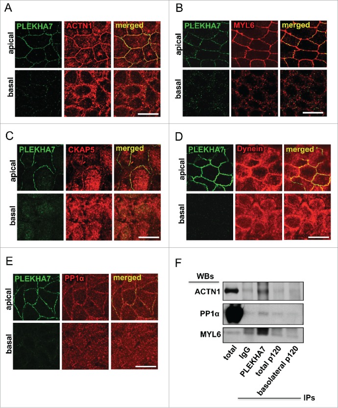
Figure 1.

PLEKHA7 associates with several cytoskeletal regulators at the ZA. Caco2 cells were grown for 21 days to polarize and subjected to IF for PLEKHA7 and co-stained for: (A) ACTN1; (B) MYL6; (C) CKAP5; (D) Dynein; and (E) PP1. In all cases, stained cells were imaged by confocal microscopy and image stacks were acquired, covering the entire polarized monolayer between the basal and the apical level. Representative x-y image stacks and merged composite x-z images are shown. Scale bars: 20 μM. (F) Western blot of the lysates from the immunoprecipitated fractions of PLEKHA7 (apical complex), total p120, and basolateral p120, isolated as previously described,15 for the markers shown. IgG is the negative immunoprecipitation control.
