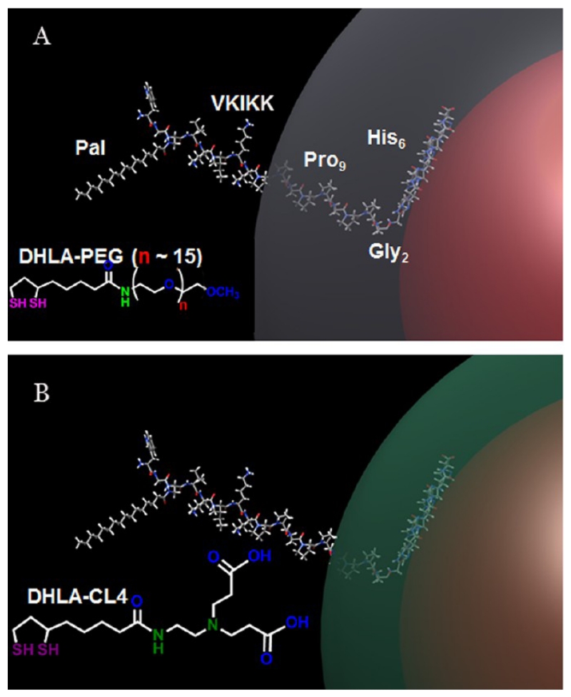
Figure 1.
Schematic depicting the QD-peptide structure. (A) Schematic representation of 625 nm QDs surface functionalized with DHLA-PEG (QD-PEG-JB577) and displaying the palmitoylated peptide (JB577) assembled by polyhistidine linkage. The 625 nm QD (diameter ~10 nm) is shown in red, and the PEG as a gray shell extending 30 Å from the QD surface. The peptide is shown in a ball and stick structure appended onto the QD by the hexahistidine sequence. Functional residues along with the position of the palmitate (Pal) group are indicated as well. The Gly2-Pro9 portion of the peptide is shown as almost completely enveloped in the PEG layer. (B) Schematic representation of the 625 QD surface functionalized with DHLA-CL4 (green shell) and binding the same peptide (QD-CL4-JB577). The structures for each respective ligand are shown inset.

