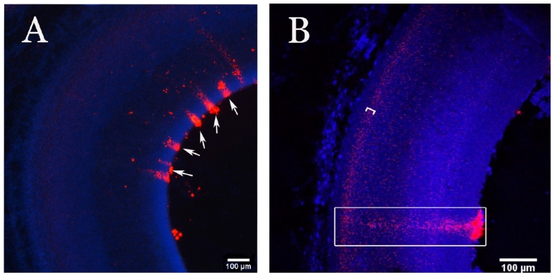
Figure 3.
Uptake and migration of the QD-peptide conjugates. Coronal 40 μm sections through midbrain of embryos injected at E4 with QD-CL4-JB577 peptide conjugates (red) and collected at E8. (A) Uptake of QDs (red) from the ventricles and evidence of long distance migration (arrows) most likely along glial tracks. (B) Higher magnification picture showing a columnar distribution of dots (box) in neuroblast migratory tracks. Another layer of cells with heavier content of QDs is indicated with a bracket, further confirming their intracellular localization.

