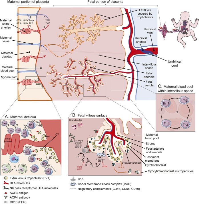
Figure 3. Immune changes in placenta of pregnant patients with NMO.
(A) Extravillous trophoblasts (EVT) invade through the uterine wall and destroy muscular wall of spiral arteries leading to increase in maternal blood flow into the intervillous space. Uterine CD16dim CD56bright natural killer cells (NKCs), which are most frequent leukocyte in decidua, can control the extent of EVT invasion into the uterine myometrium through interactions with major histocompatibility complex (MHC) molecules. Conversely, CD16bright CD56dim are mostly involved in antibody-dependent cell cytotoxicity. EVT cells that express aquaporin-4 (AQP4) antigen in AQP4 antibody–positive patients with NMO may be susceptible to be killed by cytotoxic NKCs within decidua. Furthermore, increased ratio of T-helper (Th) 1/Th2 cells and Th17/T-regulatory (Treg) cells may be involved in pathogenicity of obstetric complications in these patients. (B) Trophoblasts are the cells set in the boundary between maternal and fetal circulation of the placenta. These cells contain AQP4 water channel responsible for transferring nutrition from mother to fetus. Syncytiotrophoblasts are a special type of trophoblast made from fusion of cytotrophoblast cells. Both syncytitrophoblasts and cytotrophoblasts express AQP4 antigen and regulatory complements, though syncytitrophoblast is more exposed to maternal peripheral blood and AQP4 antibody. Binding AQP4 antibody to AQP4 antigen on syncytitrophoblast cells may activate complement cascade and membrane attack complex deposition, which further cause inflammation and necrosis in placenta. The inhibitory role of regulatory complements on complement activity in placenta of patients with neuromyelitis optica (NMO) is not completely clear. Syncytitrophoblast microparticles (STM) are released into maternal circulation during normal repairing processes or injury of trophoblasts. They cause pregnant patients with NMO to be exposed to more AQP4 antigen. Villous trophoblasts are exposed to maternal blood and lack both MHC classes (I and II). They only release an immunoregulatory nonclassical human leukocyte antigen (G) into the maternal circulation. HLA = human leukocyte antigen.

