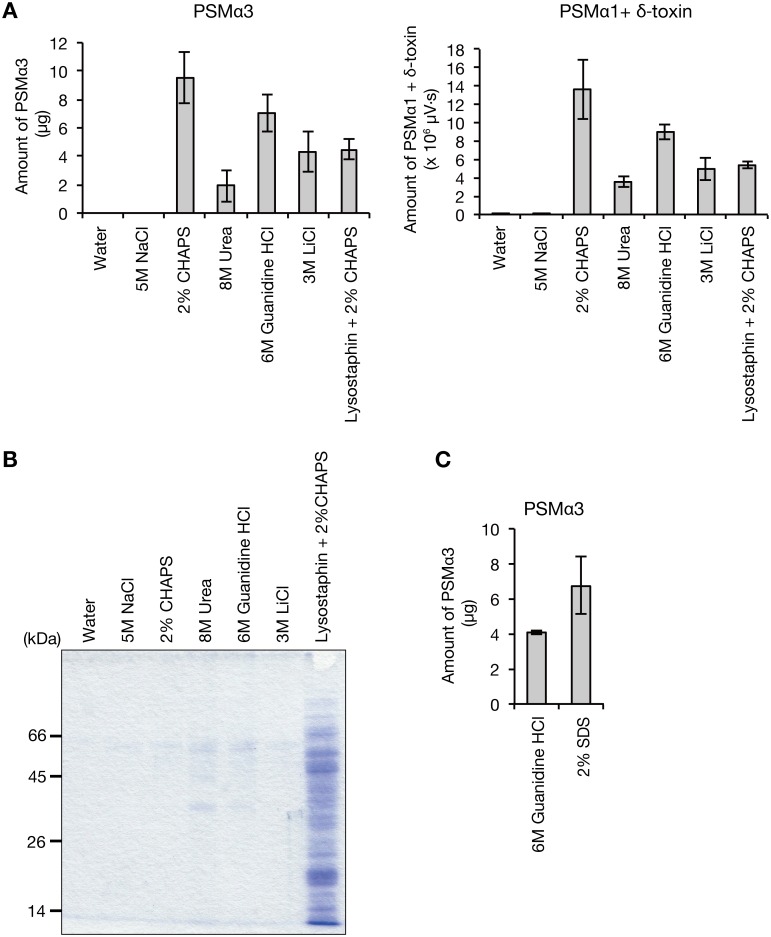
Fig 1. Presence of phenol soluble modulins on the S. aureus cell surface.
A. S. aureus Newman overnight cultured cells were washed in water, 5 M NaCl, 2% CHAPS, 8 M urea, 6 M guanidine HCl, or 3 M LiCl. In another sample, S. aureus cells were digested with lysostaphin and treated with 2% CHAPS. Samples were centrifuged and the amount of PSMα3 or PSMα1+δ-toxin in the supernatant was measured by HPLC. Vertical axis represents the amounts of PSM recovered from S. aureus cells (1.33 ml bacterial culture). Data are means±standard errors from three independent experiments. B. The centrifuged supernatants obtained in A were analyzed by SDS-PAGE. Proteins in the supernatants were precipitated with 10% trichloroacetic acid and electrophoresed on a 12.5% SDS polyacrylamide gel. The gel was stained by Coomassie brilliant blue. Each lane contains proteins from the same number of S. aureus cells (0.09 ml bacterial culture). C. S. aureus Newman overnight cultured cells were washed in 6 M guanidine HCl or 2% SDS. Samples were centrifuged and the amount of PSMα3 in the supernatant was measured by HPLC. Vertical axis represents the amount of PSMα3 recovered from S. aureus cells (1.33 ml bacterial culture). Data are means±standard errors from triplicate experiments.

