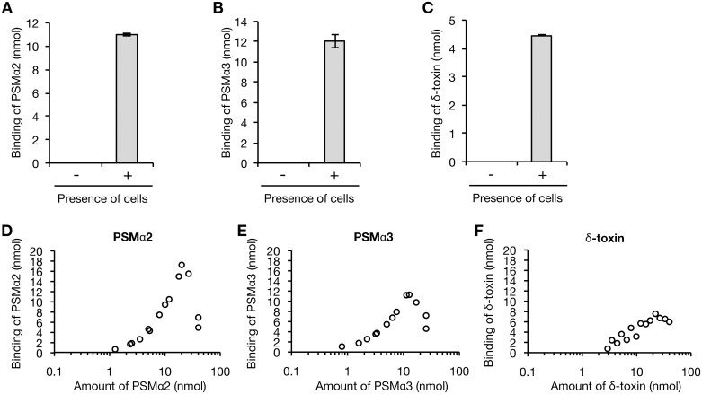
Fig 5. Binding assay of PSMs against S. aureus cell surface.
A. A-C. 10 nmol of PSMα2 (A), PSMα3 (B), or δ-toxin (C) was incubated with or without bacterial cells (3 x 10⁸ CFU) of the PSMα1-4/δ-toxin knockout strain for 30 min at 37°C. The cells were collected by centrifugation and the bound PSM was recovered by using 6 M guanidine HCl. The amount of PSM was measured by HPLC. Vertical axis represents the amount of PSM bound to S. aureus cells (3 x 10⁸ CFU). Data are means±standard errors from triplicate experiments. D-F. D-F. Dose response of PSMα2 (D), PSMα3 (E), or δ-toxin (F) to the binding to the cell surface of the PSMα1-4/δ-toxin knockout strain was measured. The bacterial cells (3 x 10⁸ CFU) were mixed with serial dilutions of PSM solutions and incubated for 30 min at 37°C. The cells were collected and the amount of the bound PSM was measured. Vertical axis represents the amount of PSM bound to S. aureus cells (3 x 10⁸ CFU). Data from two independent experiments are presented.

