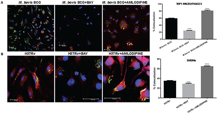
Fig 7. VGCC activation and mycobacterial infection synergistically regulate phagosome-lysosome fusion in macrophages.
PMA treated THP1 macrophages (Panel A) and mouse bone marrow derived macrophages (Panel B) were seeded on the coverslip and washed with RPMI 1640 medium and incubated in OPTIMEM medium with or without BAYK8644 for 1 h followed by infection with FM4-64 labeled M. bovis BCG (panel A) or GFP expressing M. tb H37Rv (Panel B) for 4 h. thirty minutes prior to the end of infection period, cells were incubated with 50nM of Lysotracker Green (for Panel A) or Lysotracker Green (for Panel B). At the end of incubation period cells were washed once with PBS and fixed with 4% paraformaldehyde for 1h. Following through washes, the cover slips were mounted with anti-fade containing DAPI. Confocal microscopy was performed on Leica TCS SP-8 confocal instrument, LAX Version 1.8.1.137. Bar chart in both Panel A and Panel B represents percentage of co-localization as determined by LAS AF Version 2.6.0 build 7266 of Leica Micro Systems CMS GmbH. Bars represent percentage of co-localization of the indicated groups of three independent experiments (n = 3). The stars represent the P value between unstimulated and corresponding stimulated (Bay/Amlodipine) group of that bar in each panel. The results were analyzed by one way ANOVA followed by Tukey’s post hoc multiple comparison test. * = P< 0.05; ** = P<0.01; *** = P<0.001 and **** = P<0.0001.

