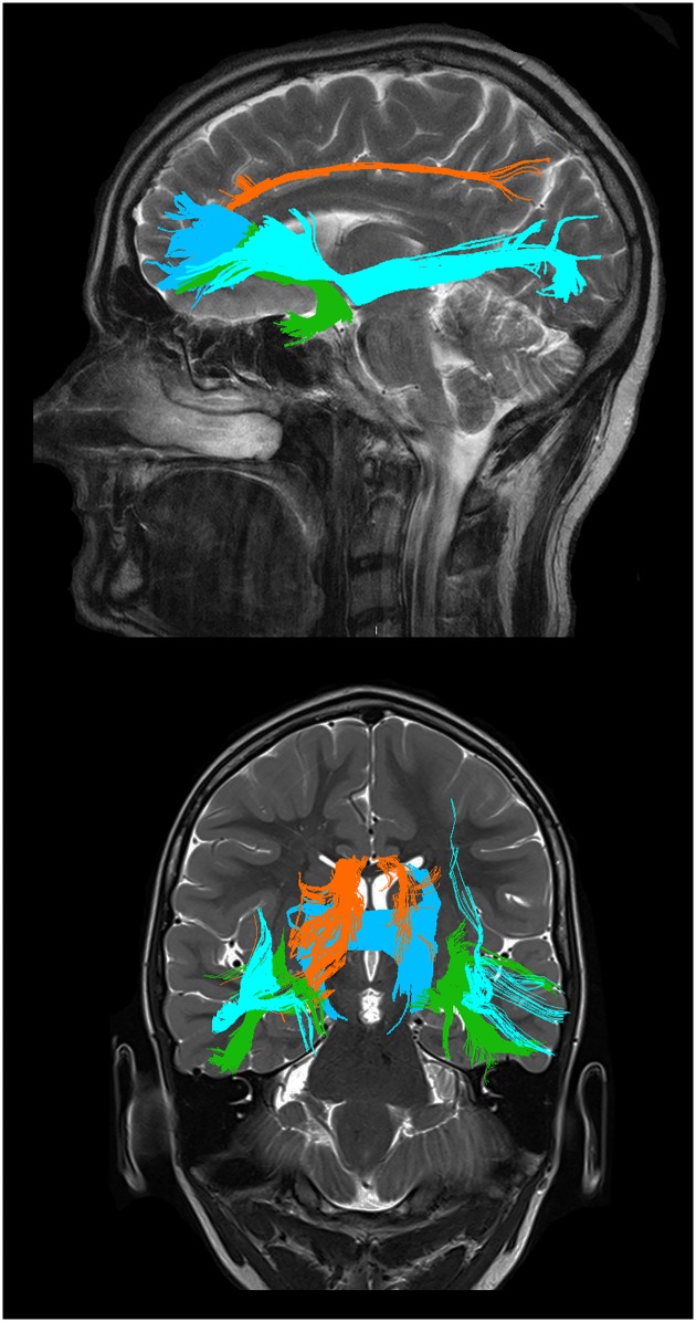
Fig 1. Graphical representations of the tracts studied.
Sagittal (upper) and coronal (lower) view. The uncinate fasciculus (green), the anterior cingulum (orange), the inferior frontooccipital fasciculus (light blue) and the forceps minor (dark blue). Tracts are from TrackVis, overlaid on high resolution images for illustrative purposes.

