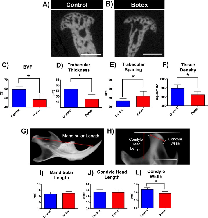
Fig 1. Reduced bone volume and density and reduced condyle width at the Botox injected side.
Coronal micro-CT images of condyles of control (A) and Botox (B) injected side masseter 4 weeks after unilateral Botox injection. Quantification of bone parameters: C) BVF—bone volume fraction, D) Trabecular Thickness, E) Trabecular Spacing, F) Tissue Density. Morphometric measurements (G-H) performed in Faxitron xray images of control and Botox injected side mandibles: I) Mandibular Lengh, J) Condyle Head Length, L) Condyle Width. Histograms (C-F, I-L) represent means ± SD for n = 13 per group. *Significant difference between control and Botox injected side (p < 0.05). Scale bar = 500μm.

