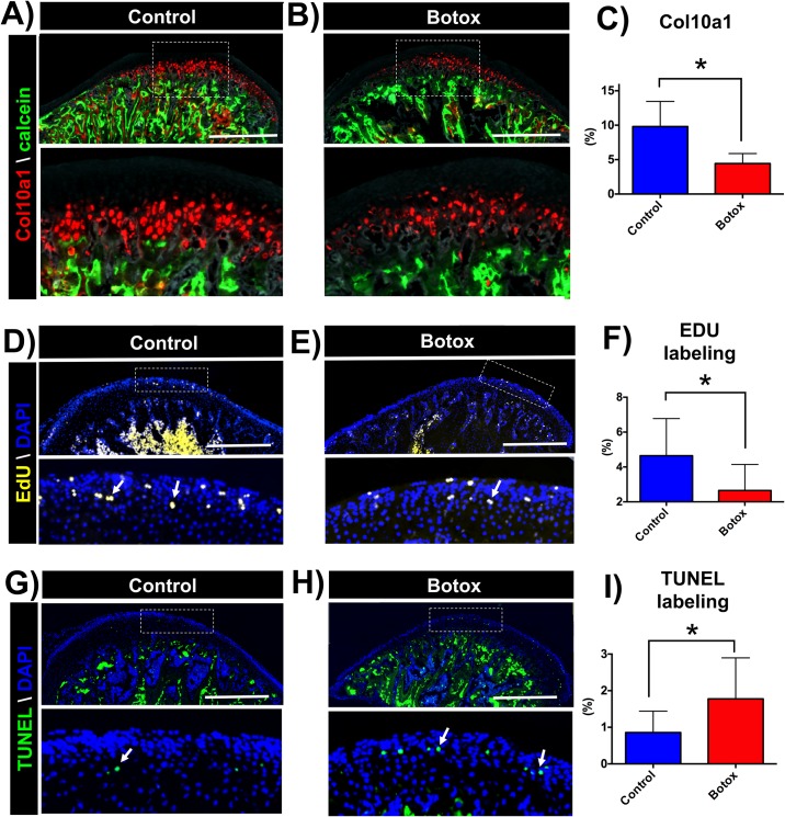
Fig 3. Reduced Col10a1 positive cells, decreased cell proliferation and increased cell apoptosis at the MCC of Botox injected side.
Representative sagittal sections of condyles of transgenic mice (Col10a1-RFPcherry), control (A) and Botox (B) injected side. C) Quantification of Col10a1 positive pixels (red) over MCC area. Sagittal sections of control (D) and Botox (E) injected side condyles stained for EdU. F) Quantification of EdU positive pixels (yellow) over DAPI positive pixels (blue) at the proliferative zone. TUNEL staining in sections of control (G) and Botox (H) injected side condyles. Histograms (C-I) represents means ± SD for n = 7 (C-F) and n = 5 (I) per group. * Significant difference between control and Botox injected side (p < 0.05). Scale bar = 500μm.

