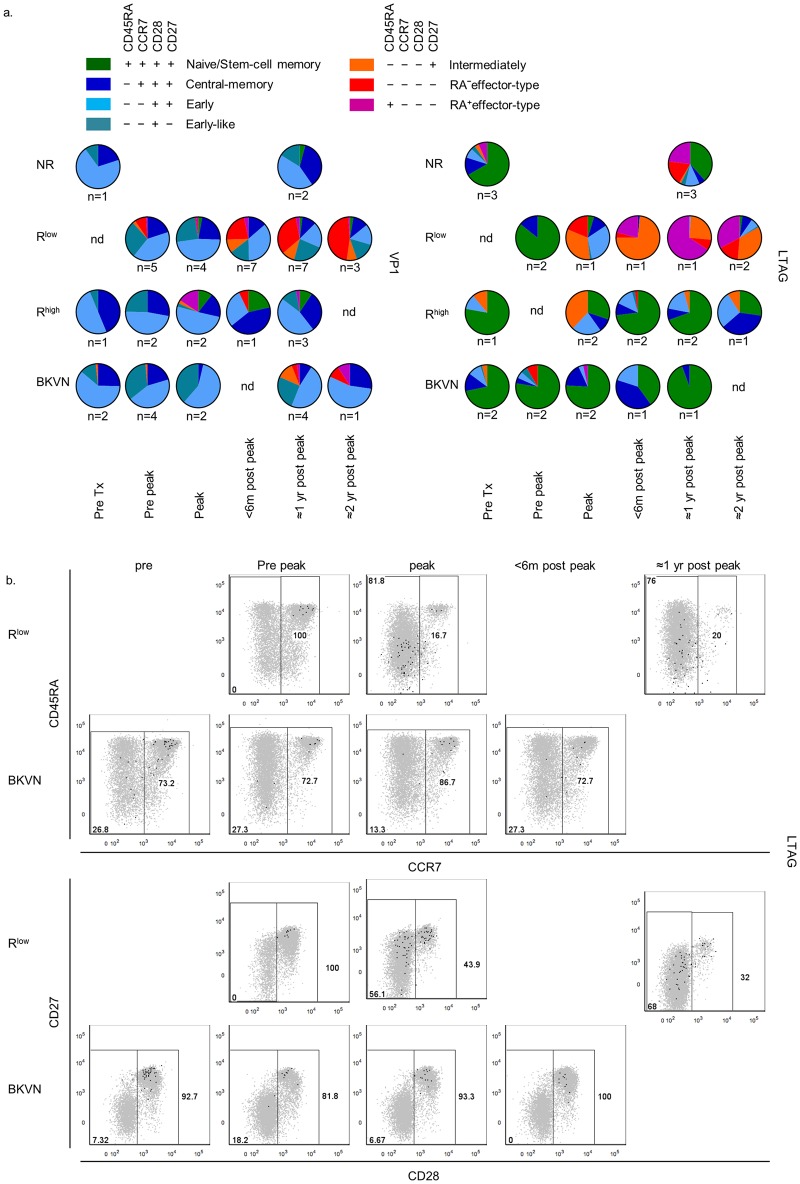
Fig 3.
(A) Pie charts depicting the distribution of the seven largest CD45RA/CCR7/CD28/CD27-defined human CD8+ T cell populations, as described previously [21], amongst VP1- (left panel) and LTAG-specific (right panel) CD8+ T cell populations detected in NR, Rlow, Rhigh and BKVN RTRs during follow-up. (B) Representative dot plot overlays showing the fluorescence intensities of CD45RA, CCR7, CD28 and CD27 with the total CD8+ T cell events shown in grey and LTAG-specific events in black from one Rlow patient (upper row) and one BKVN patient (lower row) during follow-up.

