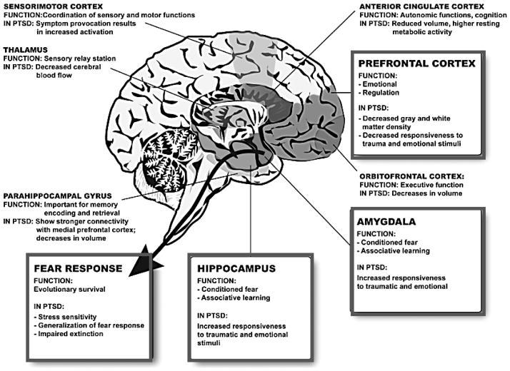
Figure 1. Brain Regions most frequently associated with Posttraumatic Stress Disorder.
This diagram of the human brain illustrates some of the most frequent brain regions associated with PTSD in two decades of work related using fMRI approaches to understand brain activation in PTSD. The prefrontal cortex (PFC) and the hippocampus have strong connections to the amygdala, which is important for conditioned fear and associative emotional learning. The PFC is involved in emotion regulation and is hypoactive in PTSD with some studies showing decreased gray matter density. The hippocampus is thought to play a role in explicit and contextual memories of traumatic events and in mediating extinction of conditioned fear. In PTSD, the hippocampus is decreased in volume. The amygdala is the most well-known area in regulating fear responses, involved in conditioned fear and recovery from fear. Hyperactivation of the amygdala to fearful cues is a robust intermediate phenotype in patients with PTSD. The end result of these neuroanatomical alterations is increased stress sensitivity, generalized fear responses and impaired extinction. Other regions including the anterior cingulate cortex, the orbitofrontal cortex, the parahippocampal gyrus, the thalamus and the sensorimotor cortex also play a secondary role in the regulation of fear and PTSD. (Figure Adapted from Mahan & Ressler, 2012).

