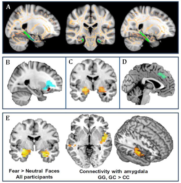
Figure 2. Genetic Associations with Structural and Functional Neural Pathways in PTSD.
Shown are examples of neuroimaging genetic approaches to identifying associations of PTSD-associated SNPs with intermediate phenotypes of neural structure and activation using structural and functional MRI. A) Differential structure of the posterior cingulum connecting hippocampus to cingulate cortex has been linked to FKBP5 SNP rs1360780. Risk allele carriers had lower fractional anisotropy in the left posterior cingulum (from Fani et al., 2014). B) The risk allele carriers of rs406001 (identified in a PTSD GWAS, Xie et al., 2013) demonstrate poorer white matter integrity in the uncinated fasciculus, connecting medial prefrontal with temporal lobe regions, including the amygdala (from Almli et al., 2014b). C) Carriers of the ADCYAP1R1 SNP rs2267735 risk allele show greater activation in amygdala to fearful faces compared to the nonrisk group (from Stevens et al., 2014). D) The number of risk alleles of SNP rs717947 (identified in a GWAS for PTSD) negatively correlated with dmPFC activity to fearful faces compared to neutral faces (from Almli et al., 2015). E) The OPRL1 gene SNP rs6010719 was found to be associated with PTSD and intermediate phenotypes of PTSD. With fMRI, enhanced bilateral amygdala activation in response to fearful versus neutral face stimuli was observed in all participants, irrespective of genotype. However, when viewing fearful faces, risk allele carriers had increased functional connectivity between amygdala and right posterior insula (from Andero et al., 2013).

