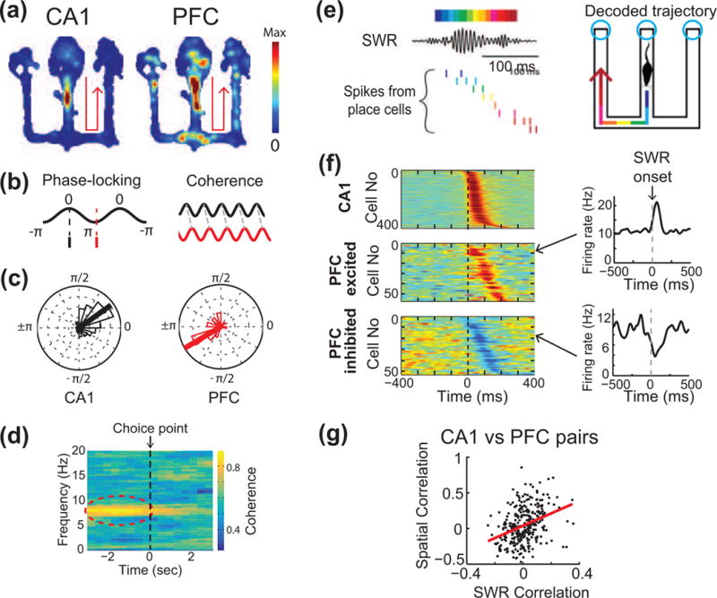
Figure 2. Theta and SWR interactions in the hippocampal-prefrontal network during behavior.

(a) Example of CA1 and PFC cells with overlapping spatial patterns of firing in a W-track spatial alternation task. Color scale indicates spiking intensity on the track. Both cells are preferentially active during outbound trajectories to right (arrows). (b) Schematic illustrating theta coupling between CA1 and PFC observed during exploratory behavior. Schematic on left illustrates phase-locking of spiking to theta oscillations; the one on right illustrates coherence, i.e. phase alignment of theta oscillations in CA1 and PFC. (c) CA1 and PFC cells are phase-locked to hippocampal theta at different characteristic phases. (d) Increase in CA1-PFC theta coherence specifically in the theta band (6–12 Hz) as animals approach the choice point in a maze. (e) Schematic illustrating replay of CA1 place cells during SWRs. Decoded CA1 activity represents spatial trajectories in the maze. SWRs are observed primarily at reward locations (cyan circles) on the W-maze. (f) Activity of CA1 and PFC cells aligned to hippocampal SWRs. PFC cells show both SWR-triggered activation and suppression of activity. (g) Reactivation of spatial information in the hippocampal-prefrontal network. CA1-PFC cell pairs with high spatial overlap are reactivated together during SWRs, while those with dissimilar patterns are suppressed. Adapted from [31**] with permission from authors.
