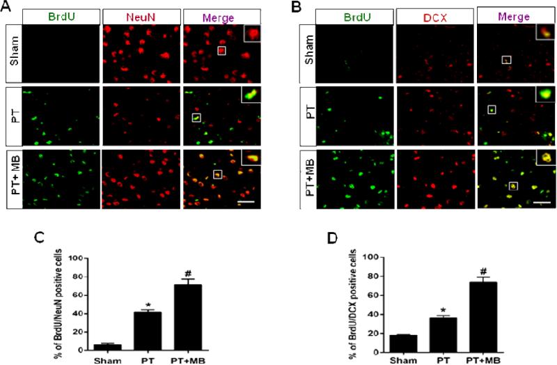
Fig. 3. Effect of MB on neurogenesis in the peri-infarct cerebral cortex on day 12 after PT stroke.
BrdU was injected on days 2 through 8. Representative confocal microscopy images of BrdU (green), NeuN (red) and DCX (red) showed that treatment with MB increases the expression and colocalization of BrdU/NeuN (A) and BrdU/DCX (B) compared to PT group. Quantitative analyses of BrdU/NeuN and BrdU/DCX are shown in (C, D). Values are expressed as mean ± SE (n=6). Magnification: 40×, scale bar: 50 μm. *P < 0.05 versus sham, #P < 0.05 versus PT group.

