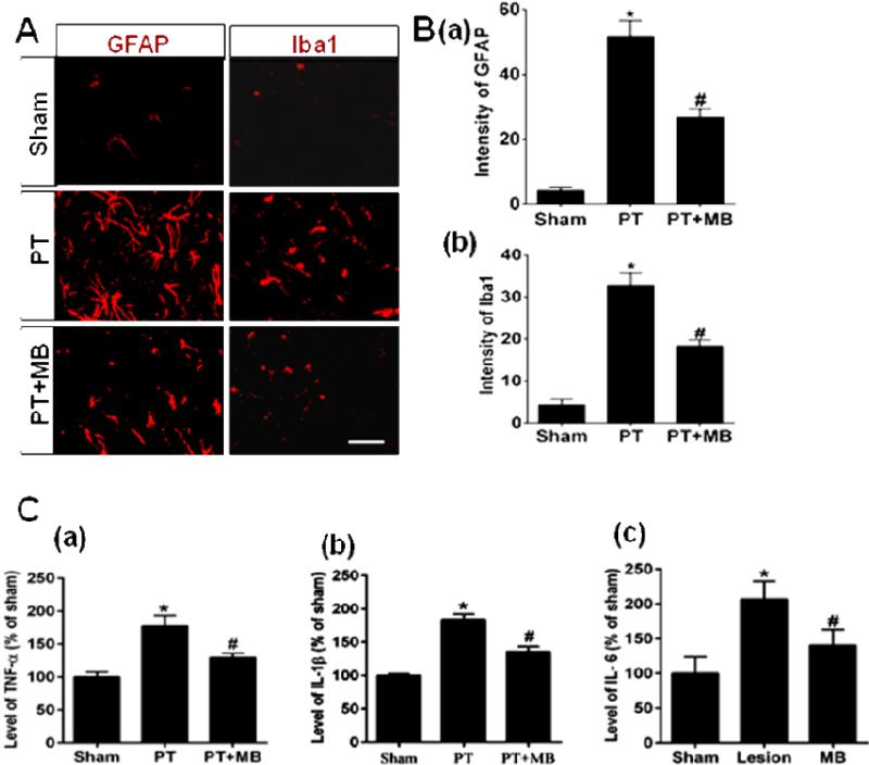
Fig. 5. Effect of MB on reactive gliosis and levels of pro-inflammatory cytokines in the infarct cortical region 12 days after PT stroke.
Distribution of microglia and astrocytes in the coronal section of peri-infarct region of cortex was immunostained with anti-Iba1 and anti-GFAP antibodies, respectively. A: Representative confocal microscopy images display the higher expression of GFAP and Iba1 in the peri-infarct area of cortex in PT group rats. C: The levels of pro-inflammatory cytokines, TNF-α, IL-1β and IL-6, were measured by ELISA assay. Note that post-treatment with MB significantly decreased the expression of reactive glia and the levels of pro-inflammatory cytokines. Values are expressed as mean ± SE (n=6). Magnification: 40×, scale bar: 50 μm. *P < 0.05 versus sham, #P < 0.05 versus PT group.

