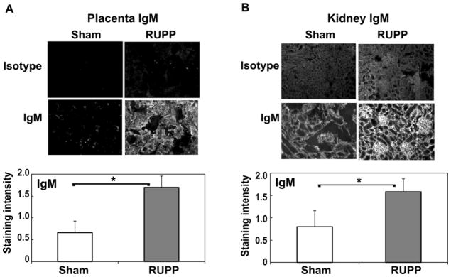
Figure 4.
IgM deposition is significantly increased in placenta (A) and kidney (B) in RUPP compared to Sham animals. Immunohistochemistry was conducted as described in Materials and Methods, and staining graded by a blinded observer from 0–3, negative to strongly positive. Representative images at 200X magnification are provided. Values for staining intensity represent the mean ± SE of scores obtained from placenta and kidney from 9–11 animals. *p<0.05 vs Sham

