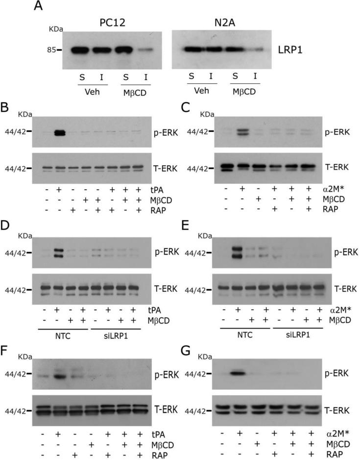
Fig. 2.
Disruption of lipid rafts blocks LRP1 signaling. (A) Immunoblot analysis comparing cell-surface LRP1 in Triton X100-soluble “S” and insoluble “I” fractions from PC12 and N2a cells after pre-treatment for 30 min with MβCD (1 mM) or vehicle. Cell-surface proteins were obtained by affinity precipitation after biotin-labeling. (B) PC12 cells were pre-treated with MβCD (+) or vehicle (−) for 30 min, and then with 150 nm RAP (+) or vehicle (−) for 30 min, followed by 12 nM EI-tPA (+) or vehicle (−) for 10 min. Immunoblot analysis was performed for phospho-ERK1/2 and total ERK1/2. (C) The study described in panel B was repeated, substituting α2M* (10 nM) for EI-tPA. (D) PC12 cells were transfected with LRP1-specific or NTC siRNA. The cells were treated with 1 mM MβCD (+) or vehicle (−) for 30 min, and then with EI-tPA (12 nM) or vehicle. (E) The study described in panel D was repeated, substituting α2M* (10 nM) for EI-tPA. (F, G) The experiments shown in panels B and C were repeated using N2a cells.

