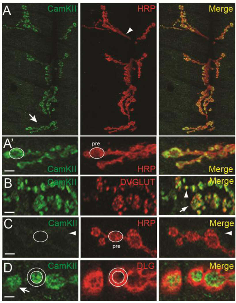
Figure 2. CamKII is enriched in presynaptic boutons.
(A) CamKII immunoreactivity in the entire synaptic arbor at muscle 6 and 7 in abdominal segment 3. Canton-S larvae were double labeled with a monoclonal antibody against CamKII (green) (Takamatsu et al., 2003) and against HRP (red). The arrow in (A) points to type I synaptic boutons shown in (A’) while the arrowhead points to the axon innervating the synaptic arbor. In (A’) an oval has been drawn around the axon terminal as defined by HRP staining and then superimposed on the green channel. Note that CamKII colocalizes with HRP within the presynaptic bouton. (B) Single confocal section of boutons of NMJs from larvae stained with monoclonal CamKII (green) and anti-DVGLUT (red) antibodies. The arrow indicates a type 1b synaptic bouton with green, red, and yellow foci. Note that there is a significant amount of overlap (arrowhead) between CamKII and DVGLUT. (C) CamKII immunoreactivity in presynaptic terminals from larvae where CamKII has been knocked down in motor neurons by RNAi (C380>CamKIIv38930). An oval has been drawn around the axon terminal as defined by HRP staining in the red channel and then superimposed onto the monoclonal CamKII (green) channel. Presynaptic CamKII immunostaining has been almost completely eliminated leaving a faint halo of immunoreactivity around the bouton border (arrow). (D) Single confocal section of boutons double stained with a polyclonal antibody against CamKII (green) (Koh et al., 1999) and against DLG (red). Two circles have been drawn to highlight the inner and outer boundaries of DLG staining and then superimposed on the green channel. Note that CamKII staining is largely restricted to the region inside the inner circle indicating enrichment with the axon terminal. Scale bar is 7.5 µm in (A) and 2.5 µm in (A’–D).

