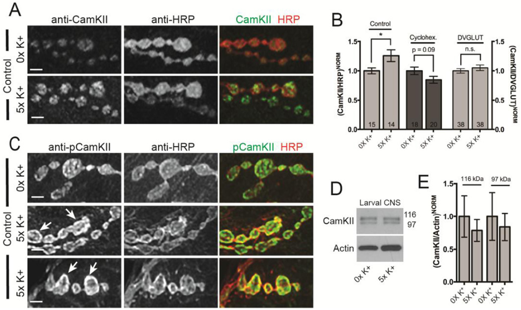
Figure 5. CamKII enrichment in presynaptic boutons is altered by synaptic activity.
(A, C) Confocal images of NMJs at muscle 6 and 7 in abdominal segment 3. Larvae were exposed to 0X and 5X high K+ spaced synaptic stimulation, fixed, and then double stained with a monoclonal antibody against CamKII or p-CamKII (green) and against HRP (red). Images in both the red and green channels were first thresholded for NMJs from the 5X high K+ stimulation group and the exact same settings used to acquire images from paired controls from the 0X high K+ group. Scale bar is 2.5 µm. (B) Quantification of the immunofluorescence from images from (A) and for NMJs double stained with antibodies against CamKII and DVGLUT. CamKII immunofluorescence in each condition is normalized to either HRP (Canton-S control and cyclohexamide-treated Canton-S control) or DVGLUT and then 0X high K+ set to 100%. * p < 0.05. The numbers of NMJs analyzed are indicated within each column. (D) Representative Western blot of CamKII in larval CNS extracts from Canton-S control animals after 0X and 5X high K+ spaced synaptic stimulation. Molecular weights for CamKII isoforms are in kDa. (E) Quantification of the levels of CamKII expression (both bands) normalized to actin from 3 independent biological replicates as shown in (C). The error bars in (B and D) indicate the SEM.

