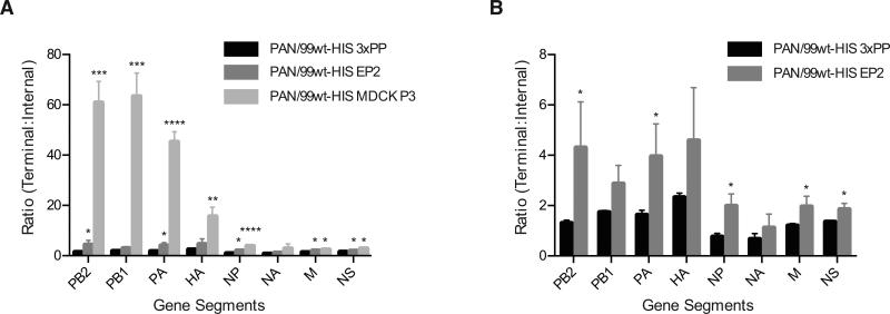
Figure 3. Defective interfering particles can be detected using droplet digital PCR.
The ratio of template copies/μl obtained with terminal and internal primers is plotted for all eight gene segments. A) Results obtained for all three virus stocks are plotted together. B) Results obtained for PAN99wt-His EP2 and triple plaque-purified (3xPP) stocks only are shown, to facilitate visualization of differences. Mean of three biological replicates is plotted and error bars indicate standard deviation. Asterisks show statistically significant differences relative to the corresponding segment of PAN99wt-His 3xPP virus (t-test). (* p<0.05; ** p<0.01; *** p<0.001; ****p<0.0001)

