Figure 5.
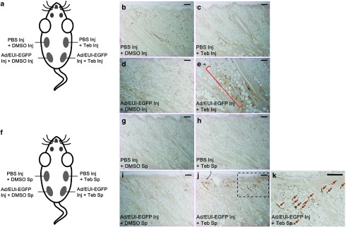
Local transgene induction in rat dermal tissues by tebufenozide treatment. In total, 20 μl of adenoviral particles (107 virus particles per μl) was delivered intradermally into rat skin. (a) A schematic diagram of EGFP transgene activation in rat skin by intradermal tebufenozide injection (Inj). After 24 hours of infection by injecting virus particles prepared in phosphate-buffered saline (PBS) or PBS alone, 20 μl of tebufenozide (100 mmol/l, dissolved in dimethylsulfoxide (DMSO)) or DMSO was administered by intradermal injection and samples were collected 48 hours later. (b) A representative tissue sample injected with PBS and DMSO sequentially. (c) A representative PBS- and tebufenozide-injected sample. (d) A virus-infected sample with subsequent injection of DMSO. (e) Cells infected with the adenoviral vector encoding the EGFP transgene (Adeno-EGFP) were stimulated by intradermal injection of tebufenozide (brown dots in a red square bracket). a–e A single animal was used for the direct comparison of the treatments. (f) A schematic drawing of EGFP transgene stimulation in rat dermis by the direct spreading (Sp) of 20 μl of tebufenozide (100 mmol/l, dissolved in DMSO) on infected skin. After 24 hours of infection by injecting (Inj) virus particles or PBS, the local infected tissues were treated with tebufenozide or DMSO for 48 hours. (g) A representative tissue sample injected with PBS and then treated with DMSO on the skin. (h) A sample injected with PBS and then treated with tebufenozide. (i) Virus-infected tissue treated with DMSO. e EGFP expression was induced in virus-infected dermis by application of tebufenozide to the skin (brown dots). (k) A magnified image of the dotted box in (j). Arrows represent tebufenozide-responsive cells. f–k A single animal was used for the direct comparison of the treatments. Integument is positioned at the top of the images. Teb, tebufenozide. Scale bar = 200 μm.

